Sonoviewer is an open access web application developed as an extension of the Mulrecon web-based viewer. The developed viewer enables volume visualization of conventional 2D ultrasound scans.
Users load an ultrasound video sweep from which a volumetric dataset is generated.
This is achieved through custom JavaScripts which extract 2D image data from the video clips.
The generated dataset can be explored as in a typical DICOM viewer with MPR capability.
The viewer utilizes Webgl 2.0 to do GPU accelerated graphics rendering.
The viewer is compatible with major web browsers such as Chrome, Firefox, and Opera.
However, at present Webgl 2.0 is not supported by the Edge and Safari browsers, but it is expected to be in the near future.
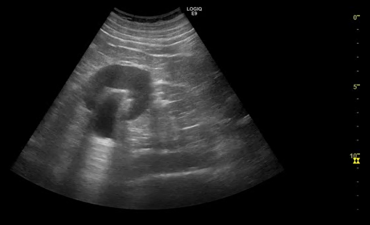
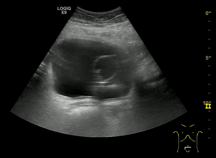
Ultrasound videos for the viewer can be obtained with a standard 2D ultrasound probe using free-hand technique. This means that the transducer needs to be moved at constant speed over the body region of interest in a regular scanning geometry in order to generate the most accurate volumetric dataset.
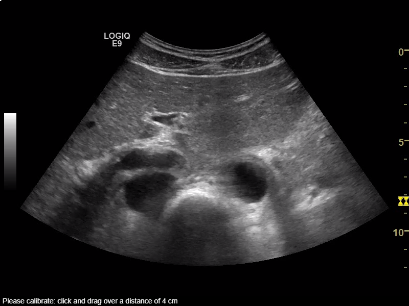
In addition the distance covered with the US sweep must be registered (Fig 1).
Link to the viewer can be found here.
The viewer can also be downloaded here and placed in a folder on a PC to be run locally or uploaded to a webserver.
In order to properly load the viewer the html file “sprayMPRGPU.html” of the viewer must be supplied with a query string “sprayMPRGPU.html?videoMode=true”.
When the viewer is opened the user is presented with a an input dialog. The workflow for loading an ultrasound video into the viewer is as follows:
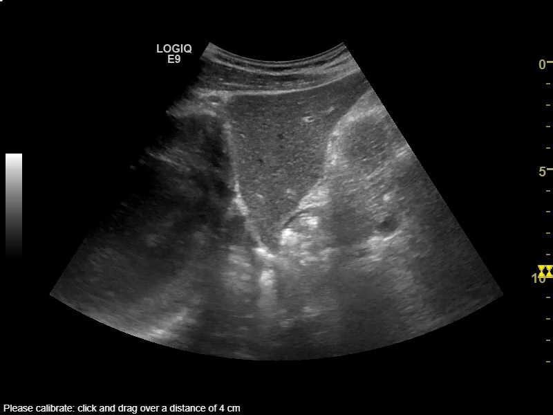
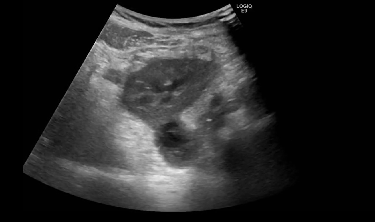
- The user select an US video file in the mp4 format for loading and specifies the approximate imaging plane and the distance between first and last frame of the video sweep (Fig 2).
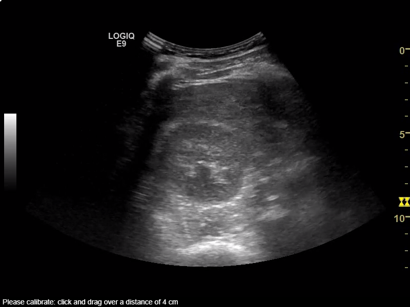
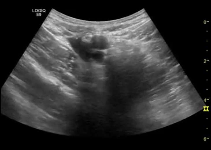
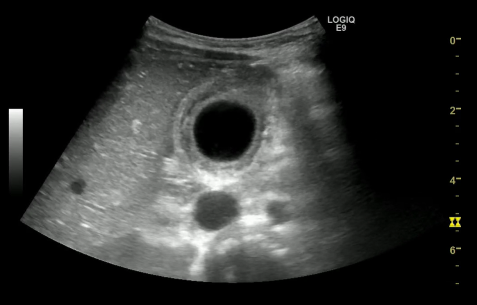
- Next, the median still image of the video is presented, and the user is prompted to place a caliper over a distance of 4 cm using the scale present on the still image (Fig 3).
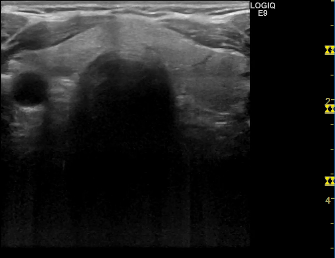
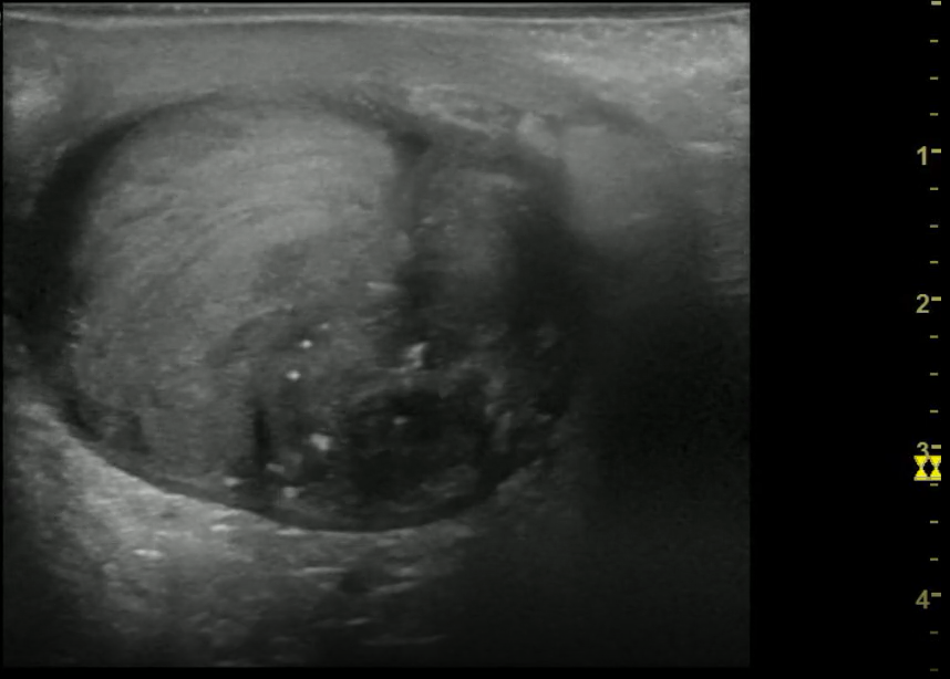
- Finally, the user is asked to draw a region of interest in the still image specifiying which part of the cine loop to be used for the volumetric dataset (Fig 4).
When these steps are completed the user is presented with a PACS-like user interface with which the dataset can interactively be explored (Fig 5).
Additionally, to enable rapid case sharing, a version of Sonoviewer connected to a web-server has been built.
This version allows upload of videos through the application which can be referenced with the Get URL button. Afterwards this URL can be shared through e-mail. Videos uploaded in this way are automatically deleted after one month.
Note: when video files are loaded in Sonoviewer no data leaves the local device running the application.
If you have any comments, see any incorrect information, typos, mistakes, or anything that you think should be added to the website or Sonoviewer, please tell us.
Sample ultrasound video clips loaded into the viewer and referenced with hyperlinks can be found below.
List of cases
-
Axial sweep of the upper abdomen (Distance between first and last video frame: 17 cm; Duration 21 s)
Viewer | download (MP4)Axial sweep of the upper abdomen. ... Read more
-
Sagittal sweep liver (Distance between first and last video frame: 15 cm; Duration 17 s)
Viewer | download (MP4)By default 3 orthogonal plans are displayed in Sonoviewer. ... Read more
-
Axial-oblique sweep right kidney (Distance between first and last video frame: 12 cm; Duration 9 s)
Viewer | download (MP4)Normal right-sided kidney. ... Read more
-
Axial sweep thyroid gland (Distance between first and last video frame: 6 cm; Duration 7 s)
Viewer | download (MP4)Demonstrates reconstructions of the thyroid ... Read more
-
Axial sweep of left-sided hydronephrosis. (Distance between first and last video frame: 12 cm; Duration 4 s)
Viewer | download (MP4)Axial sweep of left-sided hydronephrosis. Reconstructed coronal view of the dilated ... Read more
-
Axial sweep of left-sided renal graft. (Distance between first and last video frame: 13 cm; Duration 8 s)
Viewer | download (MP4)This case demonstrates thickening of the renal ... Read more
-
Axial sweep left thigh (Distance between first and last video frame: 8 cm; Duration 6 s)
Viewer | download (MP4)Case showing the blood vessels ... Read more
-
Axial sweep left thigh (Distance between first and last video frame: 7.5 cm; Duration 11 s)
Viewer | download (MP4)Ultrasound of the scrotum ... Read more
-
Axial sweep of the urinary bladder (Distance between first and last video frame: 10 cm; Duration 5 s)
Viewer | download (MP4)Ultrasound of the urinary bladder ... Read more
-
Axial sweep of the gallbladder (Distance between first and last video frame: 8 cm; Duration 6 s)
Viewer | download (MP4)Ultrasound of the gallbladder ... Read more